Understanding the cranial bones is crucial for grasping how our skull supports, protects, and adapts throughout life. In this post, we’ll dive deep into the structure and development of the cranial bones, explore what the cranial bones do (i.e., their functions), and answer some frequently asked questions in a user‑friendly way.
Overview of Cranial Bones Structure
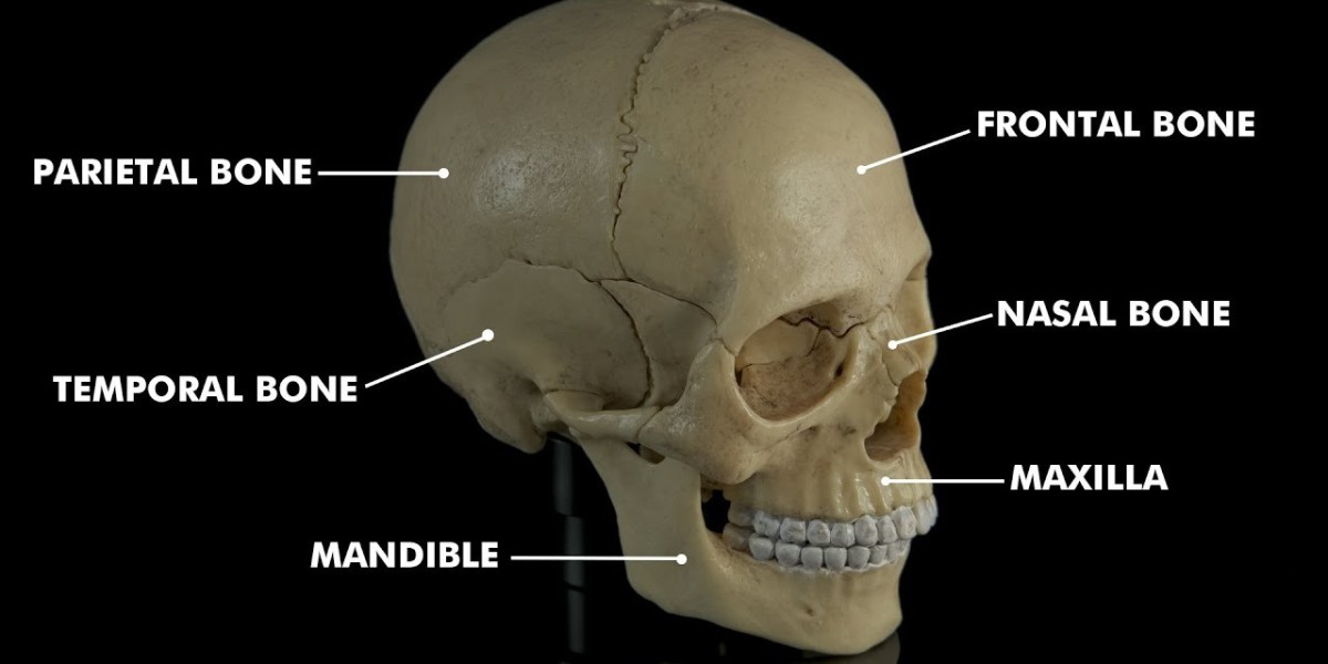
The term cranial bones refers to the set of bones that make up the skull’s vault and base, enclosing the brain and forming important bony architecture. These bones include the frontal, parietal, occipital, temporal, sphenoid, and ethmoid bones.
The Neurocranium vs. Viscerocranium
In the structure of the skull, the neurocranium (which includes many of the cranial bones) houses the brain, while the viscerocranium forms the facial skeleton. The cranial bones of the neurocranium are specially adapted to protect the brain and provide attachment sites for muscles and membranes.
Sutures, Fontanelles Bone Interfaces
The cranial bones are not rigidly fused at birth; they are connected via sutures (fibrous joints) and fontanelles (soft spots) which allow for growth and deformation during birth and infancy. These features are essential to the structure of cranial bones because they allow flexibility and expansion of the skull as the brain grows.
Flat Bones, Curved Bones Bone Plates
Many of the cranial bones (especially the vault bones, like the parietal and frontal) are relatively flat and thin, designed to cover and protect the brain, while others (like the sphenoid, temporal, or occipital) have more complex shapes. Such variation in the structure of cranial bones reflects their different mechanical and protective roles.
Development of the Cranial Bones
The development of the cranial bones is a fascinating process that begins early in embryogenesis and extends into postnatal life. It involves multiple origins, ossification types, and growth patterns.
Origins – Neural Crest Mesoderm
The cranial bones derive from two main embryonic sources: neural crest cells and paraxial mesoderm. For example, the anterior parts of the skull (viscerocranium) are largely neural crest‑derived, while the base of the skull (chondrocranium) is mesoderm derived. Understanding these origins is key to understanding variations in the cranial bones and their development.
Ossification Processes in Cranial Bones
Cranial bones develop via two main types of ossification: intramembranous ossification (common in the vault bones) and endochondral ossification (common in the skull base bones). For instance, the frontal and parietal bones form through intramembranous ossification, while the occipital base and sphenoid often form via endochondral ossification. These processes shape the final form of each cranial bone.
Postnatal Growth Suture Closure
After birth, the cranial bones continue to grow in response to brain growth; sutures remain open to allow expansion of the cranial vault. Premature fusion of sutures (craniosynostosis) can alter the shape and growth of the skull and thus the cranial bones. Therefore, the timing of suture closure is a critical part of cranial bones development.
Cranial Bones Functions
Understanding the cranial bones functions helps us appreciate why the structure and development of these bones matter. The cranial bones serve multiple roles in protection, support, and physiology.
Protection of the Brain and Sensory Organs
One of the primary cranial bones functions is to protect the brain, as well as sensory organs such as the eyes and ears. The skull’s vault and base (comprised of cranial bones) form a rigid yet somewhat flexible shell around the brain cavity. Without properly formed cranial bones, the brain and senses are more vulnerable to injury and abnormal development.
Formation of the Cranial Cavity and Supporting Membranes
The cranial bones define and maintain the shape of the cranial cavity. They provide surfaces for the dura mater and other meninges, as well as for blood vessels. The cranial bones functions include serving as attachment points for membranes and muscles, and facilitating nutrient and vascular supply.
Accommodating Growth and Structural Changes
Another key function of the cranial bones is to adapt to growth early in life, the brain grows rapidly and the skull must expand. The cranial bones functions therefore include allowing for expansions and sometimes deformations (through sutures and fontanelles) without immediate fusion. This flexibility ensures healthy brain development.
FAQs
Q1: At what age do the cranial bones fully fuse?
A1: The cranial bones do not all fuse at the same time. While major sutures begin to fuse in early childhood, full ossification and fusion may continue into adolescence or even early adulthood.
Q2: What happens when cranial bones fuse too early?
A2: When the cranial bones fuse prematurely (a condition called craniosynostosis), it can restrict skull growth, alter the shape of the head, and potentially impact brain development and intracranial pressure. Early diagnosis and treatment are important.
Q3: How many cranial bones are there and what are their names?
A3: In humans, there are eight major cranial bones: the frontal bone, two parietal bones, two temporal bones, one occipital bone, one sphenoid bone, and one ethmoid bone. These together form the neurocranium portion of the skull.
Conclusion
By exploring the structure, development, and functions of the cranial bones, we uncover how exquisitely designed this part of the human skeleton is. From embryonic origins to postnatal growth, and from protective roles to growth‑accommodating functions, the cranial bones are fundamental to both structure and function of the skull. A strong grasp of cranial bones structure development not only deepens our knowledge of anatomy, but also highlights why any disturbance in those processes can impact overall health. Whether you’re a student, health professional, or simply curious, appreciating the cranial bones offers insight into the marvel of human anatomy.